Monday, July 21, 2008

ALS cases among 1991 Gulf War veterans


Researchers probe geographical ties to ALS cases among 1991 Gulf War veterans

DURHAM, N.C. -- Researchers from Duke University, the University of Cincinnati (UC) and the Durham Veterans Administration Medical Center are hoping to find a geographical pattern to help explain why 1991 Gulf War veterans contracted the fatal neurological disease amyotrophic lateral sclerosis (ALS) at twice the normal rate during the decade after the conflict.

read the rest

Saturday, July 5, 2008

• Traumatic Brain Injuries


Why are blast injuries an important issue right now?
America's armed forces are sustaining attacks by rocket-propelled grenades, improvised explosive devices, and land mines almost daily in Iraq and Afghanistan. These injured soldiers require specialized care from providers experienced in treating traumatic brain injury
In combat, how often do blasts cause injury?
Blast injuries have become common in civilian disasters and military conflicts. It has been suggested that over 50% of injuries sustained in combat are the result of explosive munitions including bombs, grenades, land mines, missiles, and mortar/artillery shells (Coupland & Meddings, 1999).
How does a blast cause injury?
Blast injuries are injuries that result from the complex pressure wave generated by an explosion. The explosion causes an instantaneous rise in pressure over atmospheric pressure that creates a blast overpressurization wave. Primary blast injury occurs from an interaction of the overpressurization wave and the body with differences occurring from one organ system to another. Air-filled organs such as the ear, lung, and gastrointenstinal tract and organs surrounded by fluid-filled cavities such as the brain and spinal are especially susceptible to primary blast injury (Elsayed, 1997;Mayorga, 1997). The overpressurization wave dissipates quickly, causing the greatest risk of injury to those closest to the explosion.

In a blast, brain injuries can also occur by other means such as impact from blast-energized debris, the individual being physically thrown, burns and/or inhalation of gases and vapors.
What symptoms may indicate a closed head injury?
Difficulties experienced as a result of a closed-head blast injury include post concussion complaints such as decreased memory and attention/concentration, headaches, slower thinking, irritability, and/or depression.

Friday, July 4, 2008

• Hyperbaric Oxygen Therapy for TBI


A patient at the Hyperbaric Medicine Center read the recently published Honolulu Weekly article on Military Veteran Stephen Imamoto, and immediately brought the article to the attention of Helen McCracken, certified hyperbaric nurse and
Vice President of the Hyperbaric Medicine Center in Honolulu, one of 20 hyperbaric therapy centers in the United States.

The patient wanted Stephen Imamoto and others suffering from TBI (traumatic brain injury) to know about how the Hyperbaric Chamber can help them. So Helen contacted me, and after a very interesting discussion with her.

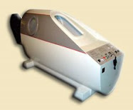
Hyperbaric Chambers help by stimulating stem cells to repair tissues with few side-effects.
Dr. Paul Harch, Emergency Medicine Specialist, Director of the LSU Health Sciences Center's Hyperbaric Medicine Fellowship Program.

Researchers Report First Successful Treatment of Chronic Traumatic Brain Injury
Posted : Thu, 11 Oct 2007 11:50:35 GMT
Author : American Association of Health Freedom
From: Earthtimes.org
ARLINGTON, Va., Oct. 11 - The American Association of Health Freedom and the International Hyperbaric Medical Association announced today that physician-researchers at the Baromedical Research Institute and Louisiana State University School of Medicine, New Orleans have reported the successful treatment of chronic traumatic brain injury (TBI). The findings, published in Brain Research (2007 Oct 12;1174:120-9), are purportedly the first-ever demonstration of improvement of chronic brain injury in animals. The treatment involved a new application and drug dosage of hyperbaric oxygen therapy.

Hyperbaric oxygen therapy has been applied to diving injuries, carbon monoxide poisoning, "flesh-eating bacteria," and chronic wounds.

Application to chronic brain injury is controversial. This demonstration is a landmark achievement that reverses 100 years of neurology and the institutionalized belief that chronic brain injury in untreatable. It has significant implications for the treatment of chronic human traumatic brain injury (TBI) such as for current U.S. military veterans.
The Navy here in Hawai'i has hyperbaric chambers available for their divers, perhaps they will throw our injured soldiers a life preserver ~ the opportunity to use their hyperbaric chamber as suggested by Dr. Paul Harch in his Testimony before Congress ~ ~ ~ ~

It has significant implications for the treatment of chronic human traumatic brain injury (TBI) such as for current U.S. military veterans.

High pressure oxygen is pumped into the body and blood giving the patient up to 20 x's the amount of normal oxygen levels.

Emerging published scientific studies are beginning to back the benefits.
VA's ongoing care for brain-injured veterans falls short (article)

Hyperbaric Medicine Program - Video (bad quality, but positive and hopeful info for soldiers)

Book Reviews:
HOPE FOR MANY "HOPELESS" DISEASES, FROM ONE OF THE FOREMOST RESEARCHERS IN THE FIELD

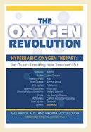
Hyperbaric oxygen therapy (HBOT) is based on an almost laughably simple idea: Oxygen can be used therapeutically for a wide range of conditions where tissues have been damaged by oxygen deprivation. Restore that oxygen, goes the logical thinking, and you can restore much of the lost function. It seems too good to be true, but Dr. Paul G. Harch’s research and clinical practice has shown that this noninvasive and painless treatment can help the tens of millions of Americans who suffer from a brain injury or disease.

It can also improve conditions in which inflammation is the culprit, such as arthritis and asthma; promote healing in infections, burns, and skin grafts, such as diabetic foot wounds; and slow the aging process [and stimulate hair growth].

For the millions of Americans suffering from these seemingly “hopeless” diseases, here finally is the handbook of hope. Inspiring and informative, The Oxygen Revolution is the definitive guide to the miracle of hyperbaric oxygen therapy, from a pioneer in the field.
Brain Injuries, Soldiers and Imaging

There's nothing more valuable than oxygen, and The Oxygen Revolution is your owner's manual May 2, 2007 David M. Freels

Paul Harch's The Oxygen Revolution simply and clearly teaches readers just what's wrong with the healthcare system. Oxygen is the most "medically necessary" substance there is, but its inherent value is all but ignored by a lazy, arrogant public health system and a corrupt, profit-driven private healthcare industry. . .

Instead of finding cures, the pharmaceutical business cycle produces drugs with outrageous price tags that only treat symptoms and not the root cause of disease. Consequently, nobody's developed a cure for any disease since polio over 50 years ago. To survive, drug companies create products that create perpetual consumers, i.e., customers. Sick people are no longer patients, and physicians are too arrogant to realize they're just pawns in the system.

The Oxygen Revolution gives us a simple biology lesson: all disease processes begin with some form of oxygen deprivation. Then Harch shows us how many, many ills of the system can be cured if patients are just given a little more oxygen.

Hyperbaric Oxygen Therapy (HBOT) can save us millions of heartaches and billions of dollars. Read The Oxygen Revolution, and you'll understand how HBOT can save our very lives and the lives of our loved ones.

David Freels, MedicaidforHBOT.com

Useful for Crush Injury and Explosion Injury


Wednesday, July 2, 2008

• DoD and VA Joint HBOT Proposal for Veterans


Proposal
Department of Defense Brain Injury Rescue & Rehabilitation Project (DoD-BIRR)

Experimental Study of Hyperbaric Oxygen Therapy
in the Treatment of Residual Effects of Traumatic Brain Injury
in United States Military Iraqi War Veterans


Paul G. Harch, M.D. • LSU School of Medicine, New Orleans

INTRODUCTION:
The current military conflict in Iraq has resulted in a very high proportion of traumatic brain injuries due to body armor and the types of munitions that are generating casualties. Statistics suggest as high as 80% of injured surviving veterans suffer from the residual neurological and cognitive effects of traumatic brain injury. There is no accepted effective treatment for these chronic effects. In the past 30 years an increasing body of scientific literature has suggested that this condition is treatable with low pressure hyperbaric oxygen therapy (HBOT). Recently, HBOT has been shown to improve cognitive function and improve vascular density in an animal model of chronic traumatic brain injury (Harch, 2001; manuscript submitted). This is the first such demonstration in the history of science and reinforces the clinical reports of effectiveness of HBOT.

PURPOSE: To conduct a rigorous study of HBOT in United States Military Iraqi war veterans with chronic traumatic brain injury and test the ability of HBOT to improve neurological, cognitive, emotional, and behavioral function.

DESIGN: Multi-center randomized prospective controlled crossover trial. All veterans enrolled in the study will receive both HBOT and participate as a control subject.

METHODS: The study will recruit 400 U.S. military Iraqi war veterans who have sustained a remote traumatic brain injury and are having cognitive and neurological dysfunction 12 months after their injury. Subjects will be identified by their VA or private physicians, enrolled in the study, and complete a neuropsychological test battery. If the neuropsychological test battery is positive for cognitive deficits the patient will undergo a neurological physical exam and randomization to HBOT or no HBOT. All veterans will complete a series of Quality of Life questionnaires and discontinue any other therapies. A subset of the veterans will also undergo the sequence of SPECT brain blood flow imaging, followed by a single HBOT or sham pressurization and then repeat SPECT at centers equipped to do SPECT. The HBOT group will receive 1.5ATA/60 minute HBOT’s twice/day, 5 days/week for 40 treatments. The control group will not receive HBOT. One month after the 40th HBOT both the HBOT and control groups will have a repeat neuropsychological test battery, answer the QOL questionnaires, and the subset with SPECT will have repeat SPECT brain blood flow imaging.

The two groups will then crossover: the HBOT group will become the control group and the control group will receive HBOT. One month after the 40th HBOT to the previous control group both groups will have repeat neuropsychological testing, QOL questionnaires, and the SPECT subset will have repeat SPECT brain imaging. Both groups will be followed for an additional six months at which time neuropsychological function, QOL, and return to work or previous level of function will be assessed. In addition, the SPECT subset will receive a final SPECT brain scan.

ANALYSIS: The neuropsychological and QOL data will be statistically analyzed by comparing pre/post test score differences for individuals as they complete each arm of the study. In addition, HBOT and control groups will be compared. SPECT brain imaging will be analyzed by standard methods, including region analysis and statistical parametric mapping. Study results will be published and announced.

COST/TIME ESTIMATES:
Each veteran will take one year to complete the study. Total cost is approximately $25,000/veteran.


Friday, March 21, 2008

ALS cases among 1991 Gulf War veterans


Researchers probe geographical ties to ALS cases among 1991 Gulf War veterans

DURHAM, N.C. -- Researchers from Duke University, the University of Cincinnati (UC) and the Durham Veterans Administration Medical Center are hoping to find a geographical pattern to help explain why 1991 Gulf War veterans contracted the fatal neurological disease amyotrophic lateral sclerosis (ALS) at twice the normal rate during the decade after the conflict.

. . . By layering military records of troop locations onto Gulf-area maps, "we've found there were some areas of service where there appears to be an elevated risk," said Marie Lynn Miranda, an associate professor at Duke's Nicholas School of the Environment whose group uses geographic information systems (GIS) to study environmental health problems.

Also known as Lou Gehrig's Disease because it crippled and ultimately killed that baseball great in 1941, ALS causes cellular degeneration in the central nervous system. Its cause is unknown.

"There are no reports on the occurrence of ALS among veterans of other conflicts," the researchers wrote. "There is only a single report that suggests ALS may arise from environmental exposures associated with military service, per se." The cases assessed by Miranda and her colleagues occurred within a group of people who are expected to be at low risk for ALS, because they're mostly under the age of 45.Miranda is the first author of a report on an initial analysis now published online in the research journal NeuroToxicology. The work was funded by the Department of Veterans Affairs Cooperative Studies Program.

The report's senior author is Ronnie Horner, professor and director of the department of public health at Cincinnati, who led research that first documented twice-normal ALS rates among vets of the first Persian Gulf War in an article published in the September 2003 issue of the journal Neurology.

Horner's group is now assessing possible exposures vets might have had in the Gulf region that could explain the higher ALS rates its 2003 study found.

"As one of the largest contemporary set of cases, it presents a real opportunity to identify clues as to the cause of ALS not only for veterans of the first Gulf War but, perhaps, for ALS generally," Horner said. UC researchers are coordinating their investigations with those of researchers at the Durham, N.C. Veterans Medical Center and nearby Duke Medical Center.

Another UC-led study, published in the July 2008 issue of the journal Neuroepidemiology, found that the risk for developing ALS has now decreased among 1991 Gulf War vets. That suggests that the cause or causes of the ALS had something to do with their deployment in the region between August 1990 and July 1991.

Of the 135 cases diagnosed among the vets within 11 years after the war, only three had a family history of the disease. The small numbers might indicate that there is an environmental cause for ALS, the authors added.

"In the one-year period of military operations, some deployed military personnel experienced numerous exposures to multiple, potentially neurotoxic agents," Miranda and coauthors wrote in the new report. "If the array of possible candidate environmental exposures could be reduced, it may be possible to identify or at least focus inquiry on specific potential causative agents."

To narrow down the possibilities, Miranda and fellow investigators used GIS analysis, which allows researchers to layer different kinds of information onto maps to deduce potential risks.

They began by searching Department of Veterans Affairs and Department of Defense records as well as other sources to identify military personnel diagnosed with ALS after 1991. Department of Defense data also allowed the researchers to identify the military units these veterans with ALS served in during their deployment to the Persian Gulf region.

In a separate analysis, the researchers identified troop units known to have been exposed to emissions from a munitions storage area at Khamisayah, Iraq. Those munitions were destroyed by U.S. forces in March 1991, and a United Nations commission later found many rockets there had been loaded for chemical warfare.

A previous Defense Department modeling study deduced that "some 90,000 veterans may have been exposed to low levels of nerve agent" at Khamisayah, the new report said.

The GIS mapping revealed that "there were some areas where there appeared to be an elevated risk," Miranda said. To narrow down the possibilities, she and co-investigators then used statistical methods that assess the "best guess about the likelihood that space matters" for each grid of Gulf territory, she added.

Applying those statistics, the likelihood of a spatial connection with ALS development "climbed as high as 91 percent" in some grid cells, she said, most notably in a region southeast of Khamisayah. But Miranda cautioned that she will need to do additional analyses that add "time" to "place" before she can be more specific.

For instance, the researchers will want to know whether the ALS victim's units were in the path of emissions from Khamisayah on a specific day. Miranda and her colleagues are also interested in examining environmental exposures that may be associated with smoke plumes from oil well fires.

Contact: Monte Basgall
monte.basgall@duke.edu
919-681-8057
Duke University
Source:Eurekaler